Types Of Hemangioma In Babies
-cavernous hemangioma is located under the skin in the adipose tissue. In rare cases a child may have hundreds of hemangiomas scattered over his body.

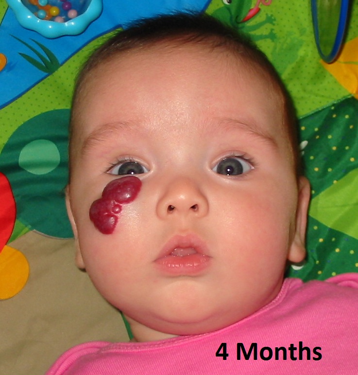
1 Superficial hemangiomas are often described as strawberry marks and appear as a bright red tumor with an irregular surface.

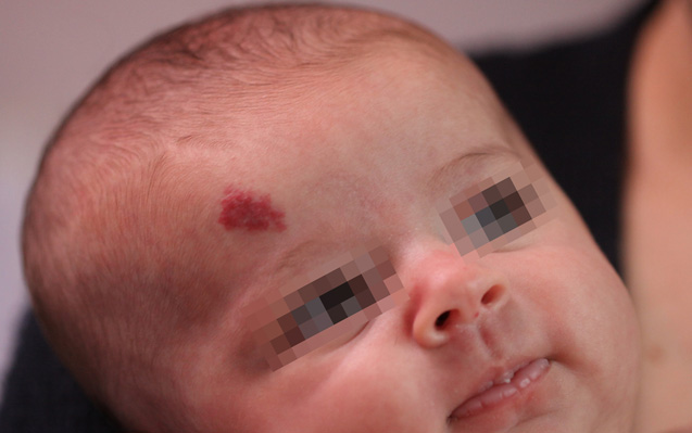
Types of hemangioma in babies. Roughly 4 to 5 of all infants get them although they are more common in Caucasians girls twins and preterm or low-birth-weight babies. Infants who have what is referred to as hemangiomatosis multiple hemangiomas are suspect for internal lesions. Strawberry birthmarks occur on the skin surface and become noticeable after a few weeks of birth.
Skin breaks down ulcerates. This type of hemangioma grows in utero is present at birth and often. -mixed hemangioma is located on the surface of the skin and subcutaneous tissues.
There may even be a hemangioma deep under his skin. The most common type of childhood vascular tumor is infantile hemangioma which is a benign tumor that usually goes away on its own. Types of hemangiomas in children.
Feeding when located on or around the mouth. Infantile hemangiomas typically go through a period of rapid growth followed by more gradual fading and flattening. Multiple superficial hemangiomas more than five can be associated with extracutaneous hemangiomas the most common being a liver hepatic hemangioma and these infants warrant ultrasound examination.
Also known as. For children who dont respond to beta blocker treatments or cant use them corticosteroids may be an option. Children may have a few skin lesions to several hundred.
Deep hemangiomas which grow under the skin in the fat may be blue purple or even skin color if they are deep enough under. Infant hemangiomas are classified based on their appearance. What are hemangiomas Congenital hemangiomas develop in utero and are present at birth.
They can be injected into the nodule or applied to the skin. All types of infantile hemangiomas are slightly warm to the touch. In this case your healthcare provider may suggest treatment with a topical medication oral medication injection laser treatment or surgery.
Internal hemangiomas referred to as visceral occur in the liver intestines airway and brain. Because malignant vascular tumors are rare in children there is not a lot of information about what treatment works best. This presents as a scarlet red lesion when it first manifests with a clean border and an irregular surface.
Infantile hemangiomas can be divided into superficial hemangiomas subcutaneous deep hemangiomas and mixed hemangiomas. Non-involuting congenital hemangioma NICH. Superficial hemangiomas which occur on the outer layers of the skin are typically bright red to purple in color.
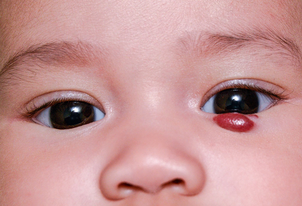
Rapidly involuting congenital hemangioma RICH. Depending on the location of the infantile hemangioma other complications can occur. Most children will have just one hemangioma.
-simple hemangioma is located on the surface of the skin. This type of hemangioma shrinks involutes without treatment and is mostly gone by the time a child is 1224 months old. Breathing when located in the airway.
Fever over 101 degrees Fahrenheit taken under the arm Skin looks open or oozes. Side effects can include poor growth and thinning of the skin. Deep hemangiomas grow under the skin making it bulge often with a blue or purple tint.
Types of Hemangioma Strawberry Hemangiomas. Rapidly involuting congenital hemangioma RICH. Deep hemangiomas are also.
A period of rapid growth often during the first year followed by a period of tumor shrinkage called involution or regression. These are symptoms of infection. Tests are used to detect find and diagnose childhood vascular tumors.
There are three general types of infantile hemangiomas. Most infant hemangiomas are capillary hemangiomas although cavernous and compound types do occur. Multiple hemangiomas most commonly affect the liver.
Girls are affected slightly more often than boys. Deep IHs present as poorly defined bluish macules that can proliferate into papules nodules or larger tumors. Diapering when in the diaper area.
The two main types of infantile hemangiomas are. Common infantile hemangiomas follow the same growth pattern. Infantile hemangiomas appear after a baby is born typically within a month.
Vision when located on or around the eye. Sometimes laser surgery can remove a small thin hemangioma or treat sores on a hemangioma. They occur deep in the skin and the vascular mass is soft and bluish with less defined borders.
Superficial hemangiomas or cutaneous in-the-skin hemangiomas grow on the skin surface. Hemangioma in the newborn may be of the following type. Very large infantile hemangiomas especially when.
When an infant has more than 3 hemangiomas an ultrasound should be done of the entire body to rule out internal lesions. Noninvoluting congenital hemangioma. The types of congenital hemangiomas are.
The section below describes the three major types of infant hemangiomas. Airway hemangiomas subglottic or diffuse hemangioma. PHACE syndrome Children with PHACE have a large hemangioma combined with other abnormalities.
Pathology Of Infantile Hemangioma Juvenile Hemangioma Cellular Hemangioma Of Infancy Dr Sampurna Roy Md
 Do You Know What A Hemangioma Is Twiniversity
Infantile Hemangioma Causes Symptoms And Treatment
 How Hemangiomas Work Howstuffworks
 Hemangioma In The Newborn Causes Symptoms And Treatment Health Care Qsota

 Infantile Hemangioma Wait Till It Goes Away On Its Own
 Infantile Hemangioma Causes Symptoms And Treatment
Infantile Hemangioma Causes Symptoms And Treatment
 Hemangioma Vascular Birthmarks Foundation

 Hemangioma Symptoms Diagnosis And Treatment
 Hemangioma Vascular Birthmarks Foundation
 Baby Birthmarks Hemangiomas Port Wine Stains And More Pampers
 Infantile Hemangiomas For Parents Norton Children S
 What Is Infantile Hemangioma Infantile Hemangioma
 Hemangioma Symptoms Diagnosis And Treatment

