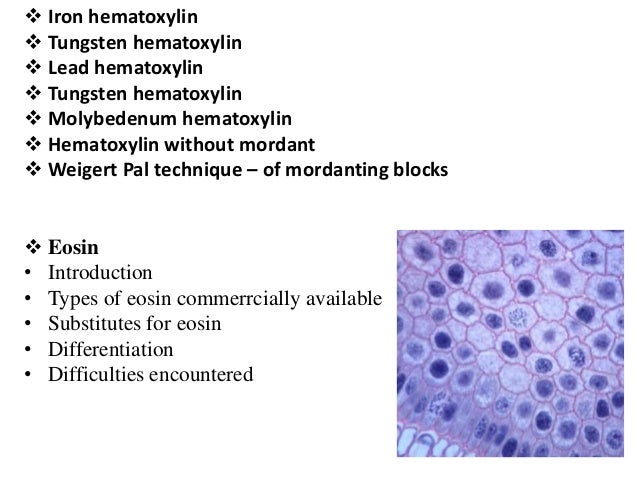
Different Types Of Hematoxylin Stains
Then stain nuclei with the alum hematoxylin Mayers to fix the tissue for about 5 minutes. Stains tissue components in various shades of blue pink red.
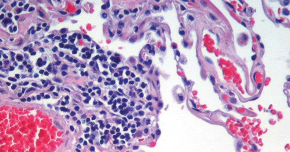 Hematoxylin And Eosin Stain H And E Stain Or He Stain
Progressive staining occurs when the hematoxylin is added to the tissue without being followed by a differentiator to remove excess dye.

Different types of hematoxylin stains. Stains nuclei blue to dark-blue. Haematoxylin requires a mordant such as iron alum which links the dye to the tissue. INTRODUCTION The word hematoxlin is drived from old Greek word Haimato blood and Xylon wood reffering to its dark red color in natural state.
Ripening of hematoxylin is a process of. It stains nuclei blue and is frequently used with eosin as a counterstain for cytoplasm. Some of them are Ehrlichs Mayers Harris Gills Delafields Coles and Carazzis haematoxylins.
Hematoxylin can be prepared in numerous ways and has a widespread applicability to tissues from different sites. Haematoxylin and its types. Haematoxylin component stains cell nuclei blue-black and shows good intracellular detail.
Based on mordants they contain there are several types of haematoxylins available. The hematoxylin stains cell nuclei blue and eosin stains the extracellular matrix and cytoplasm pink with other structures taking on different shades hues and combinations of these colors. Use as a writing and drawing ink.
The active staining chemical in ripened hematoxylin solutions. This article is concerned specifically with the chemistry of aluminum bound hematoxylin as a nuclear stain. Conversion of hematoxylin to hematein may be accomplished by the action of a number of agents.
It stains membranes and most proteins. There are typically three types of HE stains. Haematoxylin A compound used in its oxidized form haematein as a blue dye in optical microscopy particularly for staining smears and sections of animal tissue.
Eosin stains the cell cytoplasm and most connective tissue fibres in varying shades and intensities of pink orange and red. Use as a textile dye. Several older formulations such as Delafields and Ehrlichs rely on atmospheric oxygen for oxidation.
Essentially the hematoxylin component stains the cell nuclei blue-black showing good intranuclear detail while the eosin stains cell cytoplasm and most connective tissue fibers in varying shades and intensities of pink orange and red. Rinse the stain with smoothly running tap water. Alum hematoxylin Purpleblue coloration Alum-hematoxylin stains nuclei various shades of purpleblue Specific staining Non-specific staining may also occur o Cytoplasm o Mucin.
Newcomer Supply Hematoxylin Eosin HE Regressive Stain is used for screening specimens in anatomic pathology as well as for research smears touch preps and other applications. ____ of hematoxylin is necessary and may be achieved naturally. Stains the matrix of hyaline cartilage myxomatous and mucoid material pale blue.
Harris hematoxylin is used on tissue sections to stain. One of the most common staining techniques in pathology and histology. Structures that bind hematoxylin are therefore termed basophilic base loving.
Hematoxylin Hematein complexed with Al3 is the most common form of hematoxylin used for nuclear staining. Hematoxylin has a deep blue-purple color and stains nucleic acids by a complex incompletely understood reaction. 5 6 The stain shows the general layout and distribution of cells and provides a general overview of a tissue samples structure.
1 Extraction and purification. Harris hematoxylin is used on tissue sections to stain. Also use to stain metal ions eg iron lead etc.
Use as a histologic stain. Hematoxylin can be used as either a progressive or regressive stain. Progressive modified progressive and regressive.
Eosin is pink and stains proteins nonspecifically. Hematoxylin staining and counterstains 4 Hematoxylin and Hematein Common name North America Hematoxylin Hematein Common name Britain Haematoxylin Haematein Haematine Color Index number 75290 75290 Color Index name Natural black 1 Natural black 1 Ionises Acid Acid Solubility in water 3 15 Solubility in ethanol 3 7 Colour Yellow brown Dark brown. Acronym H and E stain.
Eosin is an acidic dye and the basic structures it stains are termed eosinophilic or less commonly acidophilic acid loving. The staining method involves applications of the basic dye hematoxylin which colors basophilic structures nucleic acid with blue-purple hue and alcohol based acidic Eosin Y which colors eosinophilic structures in varying shades and intensities of pink orange and red. Clean the sections to distilled water.
In these formulations hematoxylin and aluminum salts are stored in loosely capped or cotton plugged bottles to facilitate oxidation. Using the differentiator 03 acid alcohol and note the endpoint ie the correct endpoint is. 2 Use as a histologic stain.
In regressive staining tissue sections are deliberately overstained then further differentiated with dilute acid until the optimal endpoint is reached. In a typical tissue nuclei are stained blue whereas the cytoplasm and extracellular matrix have varying degrees of pink staining. Both Harris and Mayers haemoxylin formulations are aluminium-based mordant haematoxylins.
 Hematoxylin And Eosin Staining
Hematoxylin And Eosin H E Staining Principle Procedure And Interpretation
 Staining Ppt Video Online Download
 Histology Hematoxylin And Eosin Stain A Fungal Hyphae With Download Scientific Diagram
 Hematoxylin And Eosin H E Staining Of Heart Aorta And Coronary Download Scientific Diagram
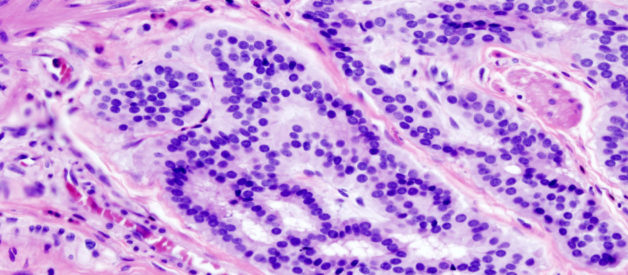 What Is Hematoxylin Eosin Stain Aladdin Creations
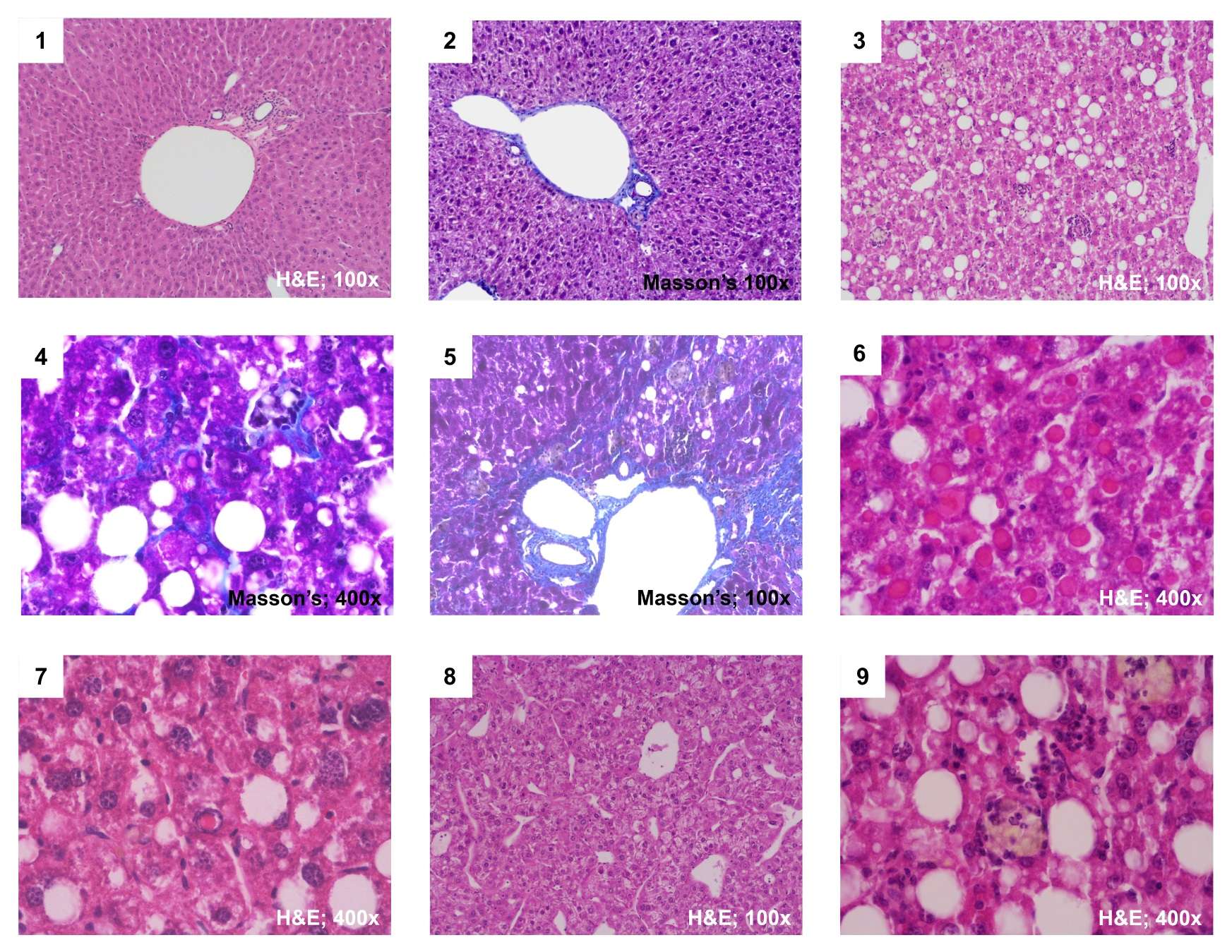 Staining The Liver Ueg United European Gastroenterology
 Bone Remodeling As Confirmed By Hematoxylin And Eosin H E Staining Download Scientific Diagram
 Histological Staining Hematoxylin Eosin Youtube
 Hematoxylin And Eosin H E Stained Liver Sections From Mice Treated Download Scientific Diagram
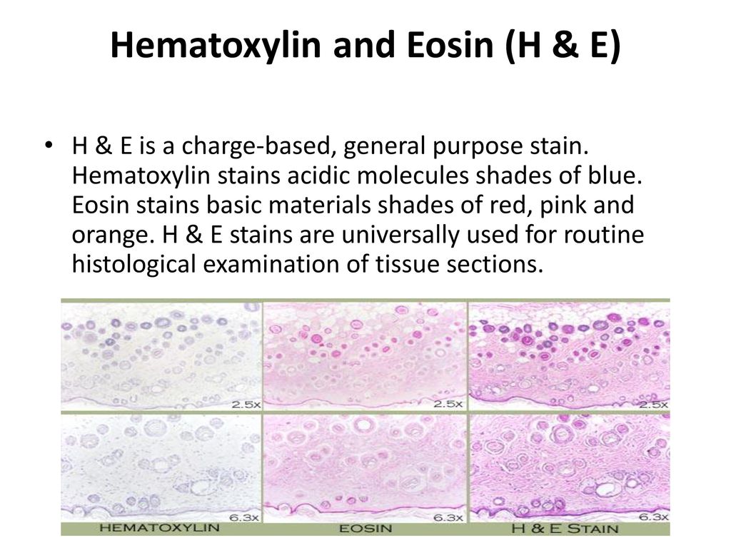 Histological Techniques Haematoxylin And Eosin Staining Ppt Download
 Colour Online Hematoxylin Eosin H E Staining Of Liver Sections For Download Scientific Diagram
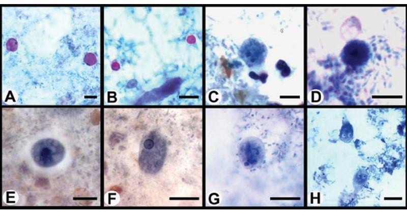 Iron Hematoxylin Staining Staining Microbe Notes
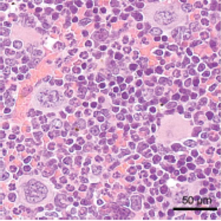 Routine And Special Histochemical Stains Histopathology Vbcf
 Hematoxylin Eosin H E Staining And Immunohistochemical Staining Of Download Scientific Diagram
Routine H E Staining Research At St Michael S Hospital
 Histology Of Mouse Pancreas Stained By Hematoxylin Eosin Normal Download Scientific Diagram
